Coronary CTA Scan -less invasive, new approach to diagnose heart disease

Less invasive, new approach to diagnose heart disease – Irish study

A clinical trial that saw high resolution scanning technology used to identify heart disease has been welcomed as a potential game-changer for cardiac treatment, being safe, minimally invasive and highly accurate.

A team at the University of Galway, in Ireland, used CT-scan imagery to pinpoint coronary artery disease and blockages as an alternative to traditional angiographs – an invasive procedure involving the puncturing of blood vessels, insertion of cables and use of dyes.

The Independent reports that the team, based at the university’s CORRIB Core Lab, analysed the images taken from patients in trial hospitals in the US and Europe.

The research was published in the European Heart Journal.

It found that the approach was 99.1% feasible, with the cardiac CT scanning offering good diagnostic accuracy without the need for invasive diagnostic catheterisation.

The trial was sponsored by the University of Galway and funded by GE Healthcare, based in Chicago, and HeartFlow, based in California.

Trial chairman Professor Patrick Serruys, established professor of interventional medicine and innovation at University of Galway, said: “The results of this trial have the potential to simplify the planning for patients undergoing heart bypass surgery.

“The trial and the central role played by the CORRIB Core Lab puts University of Galway on the front line of cardiovascular diagnosis, planning and treatment of coronary artery disease.”

The study involved 114 patients who had severe blockages in multiple vessels, limiting blood flow to their heart.

Serruys said the study offered the potential for a “monumental shift in healthcare”.

“Following the example of the surgeon, interventional cardiologists could similarly consider circumventing traditional invasive cineangiography and instead rely solely on CT scans for procedural planning,” he said.

“This approach not only alleviates the diagnostic burden in cath labs but also paves the way for transforming them into dedicated ‘interventional suites’ – ultimately enhancing patient workflows.”

A randomised trial involving more than 2 500 patients in 80 hospitals in Europe will now be undertaken.

Dr Yoshi Onuma, professor of interventional cardiology at University of Galway and the medical director of CORRIB Research Centre, said there were several benefits from the new approach.

“A catheterisation procedure is invasive and it is unpleasant for the patient,” he said. “It is also costly for the health service. While there is a minimal risk associated with the procedure, it is not entirely risk free.”

Commenting on the potential of the study, he added: “It may become a game-changer, altering the traditional relationship between GP, radiologist, cardiologist and cardio-thoracic surgeon for the benefit of the patient.”

Study details

Coronary bypass surgery guided by computed tomography in a low-risk population

Patrick Serruys, Shigetaka Kageyama, Yoshinobu Onuma et al.

Published in the European Heart Journal on 7 April 2024

Abstract

Background and aims
In patients with three-vessel disease and/or left main disease, selecting revascularisation strategy based on coronary computed tomography angiography (CCTA) has a high level of virtual agreement with treatment decisions based on invasive coronary angiography (ICA).

Methods
In this study, coronary artery bypass grafting (CABG) procedures were planned based on CCTA without knowledge of ICA. The CABG strategy was recommended by a central core laboratory assessing the anatomy and functionality of the coronary circulation. The primary feasibility endpoint was the percentage of operations performed without access to the ICA. The primary safety endpoint was graft patency on 30-day follow-up CCTA. Secondary endpoints included topographical adequacy of grafting, major adverse cardiac and cerebrovascular (MACCE), and major bleeding events at 30 days. The study was considered positive if the lower boundary of confidence intervals (CI) for feasibility was ≥75% (NCT04142021).

Results
The study enrolled 114 patients with a mean (standard deviation) anatomical SYNTAX score and Society of Thoracic Surgery score of 43.6 (15.3) and 0.81 (0.63), respectively. Unblinding ICA was required in one case yielding a feasibility of 99.1% (95% CI 95.2%–100%). The concordance and agreement in revascularization planning between the ICA- and CCTA-Heart Teams was 82.9% with a moderate kappa of 0.58 (95% CI 0.50–0.66) and between the CCTA-Heart Team and actual treatment was 83.7% with a substantial kappa of 0.61 (95% CI 0.53–0.68). The 30-day follow-up CCTA in 102 patients (91.9%) showed an anastomosis patency rate of 92.6%, while MACCE was 7.2% and major bleeding 2.7%.

Conclusions
CABG guided by CCTA is feasible and has an acceptable safety profile in a selected population of complex coronary artery disease.

Will New Lung Cancer Screening Guidelines Save More Lives?

Will New Lung Cancer Screening Guidelines Save More Lives?

Liam Davenport

DISCLOSURES

Added to Email Alert

When the American Cancer Society recently unveiled changes to its lung cancer screening guidance, the aim was to remove barriers to screening and catch more cancers in high-risk people earlier.

Although the lung cancer death rate has declined significantly over the past few decades, lung cancer remains the leading cause of cancer deaths worldwide.

Detecting lung cancer early is key to improving survival. Still, lung cancer screening rates are poor. In 2021, the American Lung Association estimated that 14 million US adults qualified for lung cancer screening, but only 5.8% received it.

Smokers or former smokers without symptoms may forgo regular screening and only receive their screening scan after symptoms emerge, explained Janani S. Reisenauer, MD, Division Chair of Thoracic Surgery at Mayo Clinic, Rochester, Minnesota. But by the time symptoms develop, the cancer is typically more advanced, and treatment options become more limited.

The goal of the new American Cancer Society guidelines, published in early November 2023, is to identify lung cancers at earlier stages when they are easier to treat.

The new guidelines, which update a 2013 version, expand the eligibility age for screening and the pool of current and former smokers who qualify for annual screening with low-dose CT. Almost 5 million more high-risk people will now qualify for regular lung cancer screening, the guideline authors estimated.

But will expanding screening help reduce deaths from lung cancer? And perhaps just as important, will the guidelines move the needle on the “disappointingly low” lung cancer screening rates up to this point?

“I definitely think it’s a step in the right direction,” said Lecia V. Sequist, MD, MPH, clinical researcher and lung cancer medical oncologist, Massachusetts General Hospital Cancer Center, Boston, Massachusetts.

The new guidelines lowered the age for annual lung cancer screening among asymptomatic former or current smokers from 55-74 years to 50-80 years. The update also now considers a high-risk person anyone with a 20-pack-year history, down from a 30-pack-year history, and removes the requirement that former smokers must have quit within 15 years to be eligible for screening.

As people age, their risk for lung cancer increases, so it makes sense to screen all former smokers regardless of when they quit, explained Kim Lori Sandler, MD, from Vanderbilt University Medical Center, Nashville, Tennessee, and cochair of the American College of Radiology’s Lung Cancer Screening Steering Committee.

“There’s really nothing magical or drastic that happens at the 15-year mark,” Sequist agreed. For “someone who quit 14 years ago versus 16 years ago, it is essentially the same risk, and so scientifically it doesn’t really make sense to impose an artificial cut-off where no change in risk exists.”

The latest evidence reviewed in the new guidelines shows that expanding the guidelines would identify more early-stage cancers and potentially save lives. The authors modeled the benefits and harms of lung cancer screening using several scenarios.

Moving the start age from 55 to 50 years would lead to a 15% reduction in lung cancer mortality in men aged 50-54 years, the model suggested.

Removing the 15-year timeline for quitting smoking would also improve outcomes. Compared with scenarios that included the 15-year quit timeline for former smokers, those that removed the limit would result in a 37.3% increase in screening exams, a 21% increase in would avert lung cancer deaths, and offer a 19% increase in life-years gained per 100,000 population.

Overall, the evidence indicates that, “if fully implemented, these recommendations have a high likelihood of significantly reducing death and suffering from lung cancer in the United States,” the guideline authors wrote.

But screening more people also comes with risks, such as more false-positive findings, which could lead to extra scans, invasive tests for tissue sampling, or even procedures for benign disease, Sandler explained. The latter “is what we really need to avoid.”

Even so, Sandler believes the current guidelines show that the benefit of screening “is great enough that it’s worth including these additional individuals.”

Guidelines Are Not Enough

But will expanding the screening criteria prompt more eligible individuals to receive their CT scans?

Simply expanding the eligibility criteria, by itself, likely won’t measurably improve screening uptake, said Paolo Boffetta, MD, MPH, of Stony Brook Cancer Center, Stony Brook, New York.

Healthcare and insurance access along with patient demand may present the most significant barriers to improving screening uptake.

The “issue is not the guideline as much as it’s the healthcare system,” said Otis W. Brawley, MD, professor of oncology at the Johns Hopkins University School of Medicine, Baltimore, Maryland.

Access to screening at hospitals with limited CT scanners and staff could present one major issue.

When Brawley worked at a large inner-city safety net hospital in Atlanta, patients with lung cancer frequently had to wait over a week to use one of the four CT scanners, he recalled. Adding to these delays, we didn’t have enough people to read the screens or enough people to do the diagnostics for those who had abnormalities, said Brawley.

To increase lung cancer screening in this context would increase the wait time for patients who do have cancer, he said.

Insurance coverage could present a roadblock for some as well. While the 2021 US Preventive Services Task Force (USPSTF) recommendations largely align with the new ones from the American Cancer Society, there’s one key difference: The USPSTF still requires former smokers to have quit within 15 years to be eligible for annual screening.

Because the USPSTF recommendations dictate insurance coverage, some former smokers — those who quit more than 15 years ago — may not qualify for coverage and would have to pay out-of-pocket for screening.

Sequist, however, had a more optimistic outlook about screening uptake.

The American Cancer Society guidelines should remove some of the stigma surrounding lung cancer screening. Most people, when asked a lot of questions about their tobacco use and history, tend to downplay it because there’s shame associated with smoking, Sequist said. The new guidelines limit the information needed to determine eligibility.

Sequist also noted that the updated American Cancer Society guideline would improve screening rates because it simplifies the eligibility criteria and makes it easier for physicians to determine who qualifies.

The issue, however, is that some of these individuals — those who quit over 15 years ago — may not have their scan covered by insurance, which could preclude lower-income individuals from getting screened.

The American Cancer Society guidelines “do not necessarily translate into a change in policy,” which is “dictated by the USPSTF and payors such as Medicare,” explained Peter Mazzone, MD, MPH, director of the Lung Cancer Program and Lung Cancer Screening Program for the Respiratory Institute, Cleveland Clinic, Cleveland, Ohio.

On the patient side, Brawley noted, “we don’t yet have a large demand” for screening.

Many current and former smokers may put off lung cancer screening or not seek it out. Some may be unaware of their eligibility, while others may fear the outcome of a scan. Even among eligible individuals who do receive an initial scan, most — more than 75% — do not return for their next scan a year later, research showed.

Enhancing patient education and launching strong marketing campaigns would be a key element to encourage more people to get their annual screening and reduce the stigma associated with lung cancer as a smoker’s disease.

“Primary care physicians are integral in ensuring all eligible patients receive appropriate screening for lung cancer,” said Steven P. Furr, MD, president of the American Academy of Family Physicians and a family physician in Jackson, Alabama. “It is imperative that family physicians encourage screening in at-risk patients and counsel them on the importance of continued screening, as well as smoking cessation, if needed.”

Two authors of the new guidelines reported financial relationships with Seno Medical Instruments, the Genentech Foundation, Crispr Therapeutics, BEAM Therapeutics, Intellia Therapeutics, Editas Medicine, Freenome, and Guardant Health.

MRI Brain illuminates the brains of ‘superagers’

MRI illuminates the brains of ‘superagers’

By Kate Madden Yee, AuntMinnie.com staff writer

July 17, 2023 — MRI shows that so-called “superagers” — people in their 80s who have the memory function of much younger individuals — have more gray matter in brain regions linked to memory function than their peers, a new study in Lancet Healthy Longevity reports.

The findings suggest that the brains of these individuals are less affected by the cognitive decline that tends to manifest as people age. Additionally, they are also more likely to have greater movement speed and lower rates of anxiety and depression, wrote a team led by doctoral candidate Marta Garo-Pascual of the Queen Sofia Foundation Alzheimer Centre at the Universidad Politécnica de Madrid in Spain.

“[Our study found] no differences in biomarkers or genetic risk factors for neurodegenerative disease … between superagers and typical older adults, suggesting superagers are resistant to age-related processes that lead to memory decline,” the investigators wrote in the study, which was published on July 13.

Whether superagers have different brains compared to typical older adults or whether they are more successful at dealing with aging has been unclear, according to Garo-Pascual.

“We are now closer to solving one of the biggest unanswered questions about superagers: whether they are truly resistant to age-related memory decline or they have coping mechanisms that help them overcome this decline better than their peers,” she said in a statement from the journal. “Our findings suggest superagers are resistant to these processes, though the precise reasons for this are still unclear. By looking further into links between superageing and movement speed we may be able to gain important insights into the mechanisms behind the preservation of memory function deep into old age.”

Previous research has found brain and lifestyle factor differences among superagers, but these studies have had small sample sizes and have not necessarily tracked changes over time. Garo-Pascual’s group sought to address the knowledge gap using data from a study called the Vallecas Project that aimed to identify early indicators of Alzheimer’s disease and included 1,213 participants.

From this total cohort, the investigators used data from 64 superagers and 55 typical older adults (all of whom were aged 79.5 years or older). Whether a person was a superager or a typical older person was based on their performance on the Free and Cued Selective Reminding Test, a tool that assesses memory function.

Study participants had up to six annual follow-up visits between 2011 and 2014 that tracked demographic and lifestyle factors. At each visit, the participants underwent brain MRI to measure gray matter volume and completed a variety of clinical tests. Researchers also took blood samples to screen for biomarkers for neurodegenerative disease and genetic risk factors for Alzheimer’s.

For the study, Garo-Pascual’s group used a machine-learning algorithm that included 89 demographic, lifestyle, and clinical predictors to identify any factors associated with superagers.

MRI showed that superagers had more gray matter — tissue key to brain function — in areas controlling memory and movement and that this gray matter tissue broke down more slowly over five years compared to the gray matter of typical older adults.

“Superagers performed better in the Timed Up and Go Test — which gauges people’s mobility — and a finger tapping test that measures fine motor function, indicating they have better mobility, agility, and balance than do typical older adults,” the team reported.

The researchers found that, compared to their peers, superagers were more active and more likely to have a musical background, greater independence in their day-to-day living, higher intelligence test scores, and lower levels of neurodegeneration biomarkers and APOE e4 (a genetic risk factor for Alzheimer’s) compared to their peers.

The AI algorithm produced similar results to the MRI data, although it was only able to distinguish superagers from typical older adults 66% of the time, which suggests that superaging could be affected by additional genetic factors, according to the investigators.

In any case, the study results add to the current knowledge base regarding superagers’ brain resilience, according to the authors.

“The brain structural signature and clinical and lifestyle factors associated with the superageing phenotype probably reflect a resistance to age-related memory decline, and these factors overlap with those associated with dementia prevention,” they concluded. “Additionally, the connection between preserved memory performance and motor function in people older than 80 provides novel insights into how to promote resistance to age-related memory loss. Taken together, the identified factors associated with superageing can inform the design of intervention trials to promote healthy ageing of episodic memory.”

Read the complete study.

MRI, CT help with planning breast cancer treatment

MRI, CT help with planning breast cancer treatment

By Amerigo Allegretto, AuntMinnie.com staff writer

August 12, 2022 — Cross-sectional supplemental imaging with MRI and CT can improve sensitivity when detecting tumors in axillary lymph nodes in breast cancer patients, a German study published August 10 in the Journal of Cancer Research and Clinical Oncology found.

Researchers led by Dr. Joachim Diessner from the Josef-Schneider-Strasse University Hospital in Würzburg found that using a combination of MRI and CT has a higher sensitivity than other combined imaging methods, allowing for safe detection of these lymph nodes at the time of diagnosis.

“Only the safe detection… enables the evaluation of the response to neoadjuvant therapy, thereby allowing access to prognosis and improving new post-neoadjuvant therapies,” Diessner and colleagues wrote.

The status of axillary lymph nodes is one of the most important prognostic factors for determining long-term survival of breast cancer patients. This includes finding out whether the nodes are infiltrated by tumors, since nodal involvement influences treatment decisions.

Supplemental imaging is used to evaluate lymph node status. While a combined method using ultrasound and mammography has been a go-to choice for radiologists, its sensitivity has a wide range depending on what previous research is cited. MRI and CT have also been explored for such supplemental imaging. However, research shows that these have lower specificity than conventional approaches.

Diessner and colleagues wanted to explore the sensitivity of pretherapeutic imaging modalities in nodal-positive breast cancer patients. They used sonography, mammography, MRI, and CT imaging. The researchers wanted to find out what further benefits could be had with cross-sectional imaging using MRI and CT for axillary staging compared with the mammography and sonography combination.

The team looked at retrospective data from 382 women who received surgery between 2014 and 2020, including 201 women who received cross-sectional imaging.

Sensitivity of combined imaging methods for axillary staging of lymph nodes in breast cancer patients
All imaging modalities 68.89%
MRI/CT 63.83%
Mammography/sonography 36.11%

As an example, the researchers noted the results of a 39-year-old patient with hormone receptor-positive, HER2-negative breast cancer. They wrote that while CT at the initial staging of the thorax and abdomen gave suspicion of lymph node infiltration, conventional imaging couldn’t clearly represent the tumor-infiltrated lymph node.

“During the period of data collection, we could detect an increasing importance of cross-sectional imaging,” the authors wrote about the MRI and CT combination.

They added that they could not prove any effect on imaging sensitivity for lymph node status when accounting for other clinical parameters such as age, intrinsic subtype, histological subtype, and body mass index.

Diessner et al noted that while prospective data would be “highly interesting,” such a study would be challenging when it comes to implementation and patient recruitment. Still, the team touted cross-sectional imaging with MRI and CT as being able to help guide treatment decisions with neoadjuvant chemotherapy with improved sensitivity.

Vogl elaborates on how best to diagnose and treat venous disease

Vogl elaborates on how best to diagnose and treat venous disease


May 4, 2021 — Venous disease is widespread and common, and it is particularly topical right now, given the potential side effects of COVID-19 vaccines. In a Q&A interview, Prof. Thomas Vogl outlines which venous disorders his team deals with in their daily clinical routine and how they diagnose and treat patients.

Vogl is head of the Institute for Diagnostic and Interventional Radiology at the University Hospital Frankfurt am Main and president of the 102nd German Radiology Congress, which takes place from 27 March to 8 November 2021. The interview was conducted by the German Röntgen Society (Deutsche Röntgengesellschaft, DRG).

Q: Which venous disorders do you deal with most frequently in your daily medical practice?

A: The human venous system and the diseases that can manifest themselves are an important field of activity for interventional radiology. Interventional radiology offers patients not only diagnostics but also minimally invasive therapeutic interventions using imaging.

Prof Dr. Thomas Vogl

Prof Dr. Thomas Vogl.

I think a lot of people don’t even know what interventional radiology can do in this area. At our institute, we very often have to deal with patients who suffer from thrombosis. These blood clot occlusions most commonly occur in the veins in the leg. In diagnostics, we clarify thromboses and possibly resulting pulmonary embolisms. The topic of thrombosis is currently very much publicized by reports on the AstraZeneca COVID-19 vaccine and sinus vein thrombosis.

Patients also come to us who suffer from varices, i.e., superficial vein enlargements that are “knotty” and clearly visible through the subcutaneous tissue. Such varices or varicose veins show up in women and men. In the case of internal vein problems, for example, we see enlarged veins that can press on nerves in the pelvic area of affected women. This condition can also occur in men, with the result that their fertility can be severely impaired. What also plays a very important role in my everyday radiology practice and in my institute are venous diseases as a result of venous malformations, which we have to evaluate.

Q: Which imaging methods do you use at your institute to diagnose venous disease?

A: The first method is ultrasound, which can determine the velocities in the venous system very precisely. The course of the vein is well documented, and it also allows the detection of superficial and medium-depth diseases of the veins. If pathologies become apparent, we use advanced imaging methods, particularly CT. This rules out a pulmonary embolism, documents the course of the vein, and determines the extent of thrombosis. With MRI, sinus vein thromboses can be diagnosed very well.

Q: On which venous disorders do you work therapeutically as an interventional radiologist?

A: In principle, interventions on the veins do not differ significantly from interventions in other diseases. Interventional radiologists treat chronically ill patients, for example by inserting port systems into the veins. In addition, when it comes to veins, we also deal with the aforementioned venous malformations and thromboses.

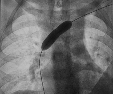
Catheter in subclavian vein

Catheter in subclavian vein. Left subclavian with tumor stenosis. Image shows large-lumen 9-mm balloon after expansion of the stenosis and before the stent is applied. Courtesy of Prof. Dr. Thomas Vogl and University Hospital Frankfurt.

A typical venous therapy is, for example, one we use on young men who have impaired fertility. The cause of this condition is dilated veins in the testicles and kidneys. Interventionally, we do it in such a way that we embolize these vein clusters and thereby restore the fertility of these — quite often — young men. We also remove foreign bodies from veins. To do this, we use instruments such as pliers, wires, and drum/barrel devices.

Q: What interventional methods are used for thrombosis?

A: In the case of thromboses, we offer the option of removing thrombi in interventional radiology. We reopen vessels that have become diseased due to chronic occlusion caused by thrombosis, and we can insert stents there to achieve outflow, for instance. We access mainly via the blood vessels — i.e., arteries or veins — with the help of catheter technology.

60-year-old male patient with severe stenosis after stent angioplasty of the right brachiocephalic vein

60-year-old male patient with severe stenosis after stent angioplasty of the right brachiocephalic vein. Images courtesy of Prof. Dr. Thomas Vogl and the DRG.

Q: Does interventional radiology also play a role in patient follow-up?

A: Chronic diseases of the veins in particular have to be checked again and again through imaging. This is especially true for venous malformations or when stents are placed in the veins. Unfortunately, diseases of the veins are usually chronic. If not handled carefully, they can lead to physical disfigurements such as varices. Above all, venous disease can have very dangerous consequences for the cardiovascular system. That is why the early detection of thromboses, tumors, or venous malformations is essential.

Editor’s note: This is an edited translation of an article published in German by the DRG on 22 April 2021. Translation by Frances Rylands-Monk. To read the original version, go to the DRG website.

Vaping injuries show particular pattern on CT

Vaping injuries show particular pattern on CT

By Kate Madden Yee, AuntMinnie.com staff writer

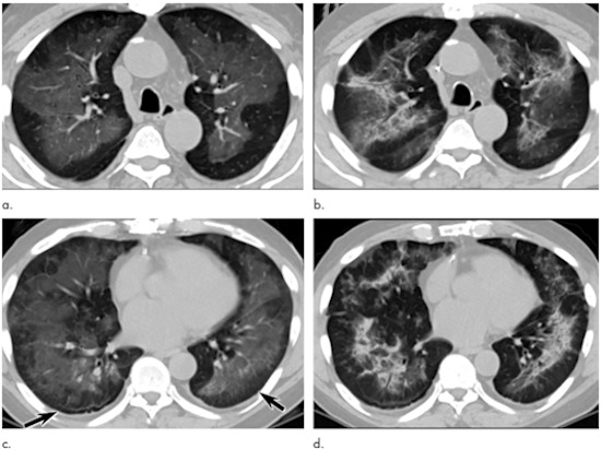
August 28, 2020 — Lung injuries caused by electronic cigarette or vaping product use-associated lung injury (EVALI) have a particular pattern on CT — namely ground-glass opacity, according to a study published August 27 in Radiology: Cardiothoracic Imaging.

The study results could help clinicians make better diagnoses of the condition and patients avoid unnecessary biopsies, wrote a team led by Dr. Michael Gotway of the Mayo Clinic in Scottsdale, AZ.

“These radiological findings will be especially beneficial to physicians to help them determine potentially less invasive treatment options,” Gotway said in a statement released by the RSNA. “We could potentially make that diagnosis noninvasively through cooperation with our clinical colleagues … [and] by alerting them to a scan pattern that is suggestive of the possibility of EVALI, we may actually save patients from having to meet a surgeon.”

Electronic cigarettes (e-cigarettes) and vaporizers heat a mix of nicotine, flavorings, and other chemicals that the user inhales, and the use of these devices has increased over the past 10 years, the RSNA said. In 2019, the U.S. Centers for Disease Control and Prevention (CDC) received the first reports of EVALI, which is characterized by “e-cigarette use or vaping within 90 days from symptom onset, associated with infiltrates at chest imaging, in the absence of an alternative explanation,” the researchers wrote. Within six months of those first reports to the CDC, 3,000 more EVALI cases were reported and 68 deaths due to the condition verified, the society said.

Gotway and colleagues investigated patterns on chest CT that might correspond to pathological findings for EVALI through a study that included 26 patients who met criteria for the condition and had undergone both CT as well as biopsy. The team classified CT findings as either ground-glass opacity or consolidation.

Electronic cigarette or vaping product use-associated lung injury in a 51-year-old man manifesting as an acute lung injury pattern at CT with subsequent organization

Electronic cigarette or vaping product use-associated lung injury in a 51-year-old man manifesting as an acute lung injury pattern at CT with subsequent organization. (a, c) Axial unenhanced CT images at presentation through (a) mid and (c) lower lungs show ground-glass opacity with subpleural sparing (arrows); this distribution was present in 45% of CT studies at presentation in our cohort (11). (b, d) Axial unenhanced CT images obtained six days later show ground-glass opacity has transitioned to consolidation and mild architectural distortion, consistent with developing organization. The patient was initially treated with antibiotics followed by corticosteroid therapy, with slow clinical improvement. Images and caption courtesy of the RSNA.

The most common CT finding among patients with EVALI also confirmed by pathology was multifocal or diffuse ground-glass opacity, identified in 96% of the study cohort — a finding even more prevalent than that of subacute hypersensitivity pneumonitis, or lung tissue inflammation, according to the authors.

“We hope to make radiologists aware that if they see diffuse lung opacities in a younger patient without clearly defined causes, then they may want to seriously consider that the patient could have a vaping injury,” Gotway said in the RSNA statement. “Patients may not readily admit that they’re using the material, but if a radiologist recognizes these patterns and then tells the clinician that it could be vaping-induced injury, then maybe they could test for a nicotine metabolite or ask the patient about this exposure.”


CT lung screening criteria overlook racial differences

CT lung screening criteria overlook racial differences

By Abraham Kim, AuntMinnie.com staff writer

June 28, 2019 — Current eligibility criteria for CT lung cancer screening from the U.S. Preventive Services Task Force (USPSTF) do not account for racial differences — leaving behind a large proportion of African American smokers who would benefit from the test, say Tennessee researchers in an article published online June 27 in JAMA Oncology.

Multiple studies have shown that African American smokers have a substantially higher risk of lung cancer than white smokers, despite smoking fewer cigarettes per day and having a lower pack-year smoking history. However, many CT lung screening guidelines set minimum age and smoking history requirements for screening without considering racial differences in smoking patterns, noted first author Melinda Aldrich, PhD, and colleagues from Vanderbilt University Medical Center.

As part of the ongoing Southern Community Cohort Study, the researchers evaluated the efficiency of using USPSTF screening guidelines in a low-income cohort consisting of predominantly African Americans (67%). The USPSTF recommends screening individuals between ages 55 and 80 with a smoking history of at least 30 pack-years.

Among 48,364 current or former smokers, the proportion of African American smokers who met the USPSTF eligibility criteria for CT lung screening was roughly half that of white smokers. This disparity in screening eligibility also applied to the subcohort of white and African American patients diagnosed with lung cancer.

The predominant reason that such a high percentage of African American smokers were deemed ineligible for CT lung screening was that they fell short of the 30 pack-year requirement, according to the authors. The proportion of African American smokers who did not meet the minimum smoking history needed for screening eligibility was roughly threefold that of white smokers.

In addition, the likelihood of developing lung cancer before reaching the minimum age requirement for screening was almost five percentage points higher for African American smokers than for white smokers (p < 0.03).

“Existing USPSTF lung cancer screening guidelines do not appear to be optimized for African American smokers and may result in a widening of racial disparities in late-stage diagnosis, potentially leading to higher mortality and worse outcomes among African American persons with lung cancer,” Aldrich and colleagues wrote. Yet simple adjustments to the USPSTF guidelines could help mitigate these disparities, they continued.

For example, the researchers showed that modifying the screening criteria to cover African American smokers with a history of 20 pack-years would increase the proportion of African Americans eligible for screening to 28.5%, compared with 17.4% for the current 30 pack-year requirement. Lowering the minimum screening age to 50 years from 55 years for African Americans would boost their eligibility even further.

What’s more, such revisions to the eligibility criteria would improve the sensitivity of CT lung screening for this cohort and narrow the gap between African American and white smokers, the authors noted.

“Current USPSTF lung cancer screening guidelines may be too conservative for African American smokers. The findings suggest that race-specific adjustment of pack-year criteria in lung cancer screening guidelines would result in more equitable screening for African American smokers at high risk for lung cancer,” they concluded.

Should NICE Guidelines Be Universally Accepted for the Evaluation of Stable Coronary Disease?


A Debate

Harvey S. Hecht; Leslee Shaw; Y.S. Chandrashekhar; Jeroen J. Bax; Jagat NarulaDISCLOSURES

Eur Heart J. 2019;40(18):1440-1453. 

Abstract and Introduction 

Abstract

The 2016 National Institute of Health and Care Excellence clinical guideline for the assessment and diagnosis of chest pain positions coronary computed tomography angiography as the first test for all stable chest pain patients without confirmed coronary artery disease and discards the previous emphasis on calculation of pre-test likelihood recommended in their 2012 edition of the guidelines. On the other hand, the American College of Cardiology Foundation/American Heart Association and the European Society of Cardiology guidelines continue to present the stress testing functional modalities as the tests of choice. The aim of this review is to present, in the form of a debate, the pros and cons of these paradigm changing recommendations, with an emphasis on literature review and projection of future needs, with conclusions to be drawn by the reader.

Introduction: Nice Guidelines for Evaluation of Stable Coronary Disease

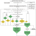
The National Institute of Health and Care Excellence (NICE) is the executive non-departmental public body of the Department of Health in the United Kingdom responsible for determination of health care policy for both the English and Welsh National Health Service. The 2016 NICE update for the Chest Pain of Recent Onset: Assessment and Diagnosis Clinical Guideline[1](Figure 1) was proposed after exhaustive review and analysis of accuracy, outcomes and cost effectiveness, and has since replaced the 2012 version.[2] The guideline positions coronary computed tomography angiography (CCTA) as the first test for all stable chest pain patients without confirmed coronary artery disease (CAD), and (interestingly) discards the previous emphasis on calculation of pre-test likelihood recommended in the 2012 edition of the guidelines. In the penultimate version, the first line investigations were stratified as invasive coronary angiography (ICA) for estimated CAD likelihood of >60–90%, functional imaging for >30–60%, and coronary artery calcium (CAC) scan for estimated CAD likelihood of >10–30%.[2] However, in patients with previously confirmed CAD, functional imaging and maximum endurance exercise treadmill testing (ETT) remain as the recommended first line tests for any change in clinical status or for timely follow-up. The NICE document has further added that fractional flow reserve (FFR) derived from computed tomography angiography (CTA) (FFRCT) should be considered as an option for patients with stable, recent onset chest pain who are offered CTA as part of the NICE pathway on chest pain, and have projected significant cost savings accruing from its utilization.[3]

Click to zoom

(Enlarge Image)

Figure 1.

National Institute of Health and Care Excellence (NICE) guideline for ‘Chest Pain of Recent Onset Assessment and Diagnosis’.2 CAD, coronary artery disease; CT, computed tomography; ECG, electrocardiogram; MR, magnetic resonance. Reprinted with the permission of the publisher from Chest Pain of Recent Onset.1

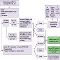
On the other hand, 2012 American College of Cardiology and American Heart Association (ACCF-AHA) guidelines for the diagnosis and management of patients with stable ischaemic heart disease[4] (Figure 2) and 2013 European Society of Cardiology (ESC) guidelines on the management of stable CAD[5] (Figure 3), rely on pre-test likelihood and offer a multiplicity of functional imaging tests as the first line diagnostic tool. The ACCF-AHA Guidelines support Class Ib recommendation for ETT, stress echocardiography (SE), and myocardial perfusion radionuclide imaging (MPI) and IIa for magnetic resonance imaging (MRI). Computed tomography (CT) angiography is reserved for patients with low-to-intermediate pretest probability of CAD who are incapable of at least moderate physical activity or have disabling comorbidity (Class IIa recommendation), and for patients with an intermediate pretest probability of CAD who can undertake at least moderate physical activity or reveal no disabling comorbidity (Class IIb recommendation). The ESC Guideline designates a Class Ib recommendation for all functional modalities, including MRI and positron emission tomography (PET). European Society of Cardiology also includes a Class IIa recommendation for CTA in patients in the lower range of intermediate pre-test probability when adequate diagnostic image quality can be expected, as an alternative to stress imaging, or after an inconclusive stress test, or for patients who have contraindications to stress testing in order to avoid otherwise necessary ICA. However, neither ACCF-AHA nor ESC documents had the benefit of the extensive CTA literature published since 2012–2013, which weighed into formulation of the NICE guidelines 2016 update vis-à-vis their departure from the conventional practices reflected in the 2012 American and 2013 European guidelines. It is not certain whether or not the next ACCF-AHA and ESC guidelines will come to similar conclusions as 2016 NICE update. However, it is important to examine the implications for the practicing cardiologists if the NICE guidelines were to be implemented universally.

Click to zoom

(Enlarge Image)

Figure 2.

2012 ACCF/AHA/ACP/AATS/PCNA/SCAI/STS guideline for the diagnosis and management of patients with stable ischaemic heart disease.4 CCTA, coronary computed tomography angiography; CMR, cardiac magnetic resonance; ECG, electrocardiogram; Echo, echocardiography; IHD, ischaemic heart disease; MI, myocardial infarction; MPI, myocardial perfusion imaging; Pharm, pharmacological; UA, unstable angina; UA/NSTEMI, unstable angina/non-ST-elevation myocardial infarction. Reprinted with the permission of Elsevier from Fihn et al.4

Click to zoom

(Enlarge Image)

Figure 3.

European Society of Cardiology 2013 non-invasive testing in patients with suspected stable coronary artery disease and an intermediate pre-test probability.5 CAD, coronary artery disease; CTA, computed tomography angiography; CMR, cardiac magnetic resonance; ECG, electrocardiogram; ICA, invasive coronary angiography; LVEF, left ventricular ejection fraction; PET, positron emission tomography; PTP, pre-test probability; SCAD, stable coronary artery disease; SPECT, single-photon emission computed tomography. Reprinted with permission of Oxford Academic from Montalescot et al.5

How calcium in coronary arteries can predict future heart health


Published Sunday 23 June 2019 By Lauren Sharkey

Fact checked by Jasmin Collier

According to one new study, having high levels of calcium in the coronary arteries could be responsible for detrimental changes to the structure of the heart.

Heart anatomy model

A new study investigates calcium and heart disease.

“Heart disease is the leading cause of death for men and women,” according to the Centers for Disease Control and Prevention (CDC).

Being able to identify people at risk is therefore a crucial public health issue.

One way to determine a person’s risk of heart diseasestroke, or heart attack is by looking at their coronary artery calcium (CAC) levels.

Calcium plays a number of roles in the body, including keeping bones healthy. However, calcium present in coronary arteries can lead to the accumulation of plaque.

Over time, this calcified substance can cause atherosclerosis, or a narrowing of the arteries. Atherosclerosis restricts blood flow and oxygen supply to vital organs, potentially resulting in a heart attack or stroke.

High cholesterol levels can indicate that a person is at risk; but scientists can also test CAC levels directly.

Using a CT scan to take numerous sectional pictures of the heart, doctors can see specks of CAC. A person’s scores tend to range from zero to over 400. The higher the score, the higher the risk of developing cardiovascular disease.

Cholesterol guidelines from 2018 recommend a CAC scan for people ages 40–75 whose risk status is “uncertain,” note the American Heart Association (AHA).

A new study, the results of which now appear in the journal Circulation: Cardiovascular Imaging, has examined the CAC scores of younger people and drawn some interesting conclusions.

Heart abnormalities

The scientists used data from almost 2,500 people to track CAC and heart structure differences between young adulthood and middle age. Women made up 57% of the group, and 52% of participants were white.

They took data from participants in the Coronary Artery Risk Development in Young Adults (CARDIA) study, which began in the 1980s with the aim of identifying young adult risk factors for cardiovascular disease.

“We looked at early adulthood to middle age because this is a window in which we can see abnormalities that might not be causing symptoms, but could later increase the risk of heart problems,” explains study co-author Dr. Henrique Turin Moreira.

The researchers compared test results from years 15 and 25 of the CARDIA study period. At the 25-year mark, the average age of the group was around 50.

When it came to their CAC results, 77% of participants had a score of zero in year 15 of the study. However, in year 25, this had dropped to 72%i

A number of factors were linked to a rise in CAC scores, including being older, being male, being black, smoking, having higher cholesterol levels, and having higher systolic blood pressure.

Middle-aged people who had higher CAC scores also showed a 9% increase in left ventricular volume and a 12% increase in left ventricular mass.

When the left ventricle changes in this way, the heart has to put more effort into pumping blood. This, in turn, leads to a thickening of the heart, which increases the risk of heart failure.

The study authors also note that these abnormalities were more significant among black people. For these people, every one-unit change in their CAC score correlated with quadruple the increase in their left ventricular mass.

Future implications

It is unclear why people exhibited such differences depending on their race. Dr. Moreira explains that it could be “due to genetic factors or perhaps greater exposure to cardiovascular risk factors that usually appear earlier” in black people.

What that do already know, however, is that black people are already more likely to develop cardiovascular disease. Although just 43% of white women and 50% of white men have cardiovascular disease, it affects 57% of black women and 60% of black men.

Further research, explains Dr. Moreira, will be needed to “examine the link between coronary artery calcium and heart health” — especially in relation to race. However, documenting the relationship between CAC and heart failure risk factors in a younger age group is significant.

“Given the burden of morbidity and mortality associated with heart failure, these are important findings,” says Dr. Salim Virani, a co-author of the AHA’s 2018 cholesterol guidelines.

“Prior studies from this cohort have also shown that a better risk factors profile in young adulthood is associated with much lower CAC and therefore, these results further highlight the importance of primordial prevention and risk factor modification in early adulthood.”